The knee joint
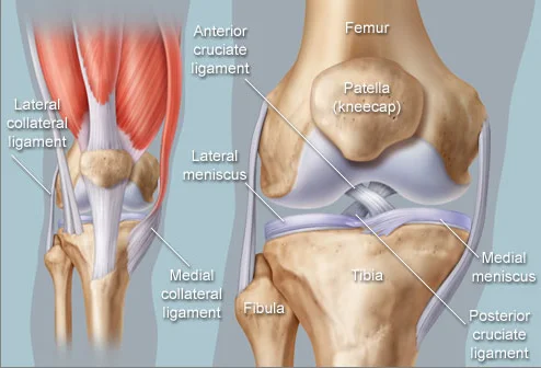
The knee joint is formed by the lower end of the thigh bone and the upper end of the shin bone (tibia). The ends of the bones are covered with a smooth layer of cartilage. The whole joint is enclosed in a fibrous capsule, lined on the inside with smooth tissue called the synovium. The synovium produces a fluid that reduces friction and wear in the joint.
Normally the joints move freely. When the cartilage is damaged by arthrosis or other causes, the joints become stiff and painful.
Arthrosis and inflammation
Arthrosis
Arthrosis is the result of wear of the articular cartilage. Normal cartilage gradually wears and thins, and the friction between the bones grows. This can go on for many years before the damage to the joint becomes noticeable.
Pain and stiffness are the usual problems of osteoarthritis. The inner part of the knee is affected most often. Earlier injuries of the knee ligaments and the menisci can predispose to the condition. Being overweight can also make arthritis more likely, or more severe.
Inflammation
Joints can become inflamed for different reasons; the most common is rheumatoid arthritis. The inflammation damages the cartilage of the joints and causes arthritis that often affects several joints. This kind of arthritis can affect people when they are relatively young and can lead to serious damage.
Treating arthritis
To treat arthritis properly, its nature has to be established. Blood tests and other examinations can help with this. Anti-inflammatory and other medicines can be very useful.
For patients with osteoarthritis it is especially important that the joints stay flexible and keep as much movement as possible. Analgesics (painkillers) can help patients keep up their daily activities. Many modern painkillers also have an anti-inflammatory effect that can help with arthritis.
Knee replacement
Total knee replacement
When the cartilage of the knee is badly worn, the treatment is surgical. In a knee replacement, the damaged bone and cartilage are replaced with metal and polymer components shaped to restore the movement and function of the knee. The new artificial knee is called an endoprosthesis.
It usually consists of two metal components fitted to the ends of the tibia and the femur, with a polymer component between them. The metal parts are made of steel, cobalt-chrome or titanium alloys. Durable, wear-resistant polyethylene (plastic) is used where the metal parts meet. The components are fixed to the bone with so-called bone cement, or without cement, directly to the bone.
Partial knee replacement
When only one part of the knee is affected by the disease, it is possible to replace only that part. The procedure is similar to a total knee replacement: parts of the femur and the tibia are replaced, with a polyethylene component between the metal ones. Recovery is usually a little shorter after this kind of operation.
Patients have to be selected carefully here. It is not unusual for a total knee replacement to be needed a few years later.
Corrective knee operations
Sometimes the state of the joint allows a so-called corrective osteotomy.
Tibial osteotomy
In this operation, called a high tibial osteotomy, a wedge of bone is taken out of the tibia to change the angle at which the knee carries weight. The heavy load is thus moved onto the relatively healthy part of the joint. The correction is held with metal implants, and intensive rehabilitation is needed after the operation. Although the procedure is not suitable for every patient, it can bring considerable relief and delay the need for a knee replacement.
Anaesthesia for knee surgery
A knee replacement can be done under general, spinal or epidural anaesthesia. A combination of techniques is often used. Your anaesthetist will suggest the best anaesthesia in your case.
General anaesthesia
The patient is asleep during the procedure, and breathing is supported by a machine. Many patients feel drowsy or very weak after a general anaesthetic. Modern medicines greatly reduce nausea and vomiting after the operation.
Spinal or epidural block
These techniques block nerve impulses, including pain, in the spinal cord. They are regional anaesthesia. The block can be placed at different levels of the spine, but the patient usually has no sensation from the waist down. Depending on the medicines used for the block, pain relief can last several hours after the operation, so fewer painkillers are needed. Because these blocks cause a temporary loss of sensation below the waist, a urinary catheter (a tube into the bladder to drain urine) is often needed.
Possible complications
Every surgical procedure carries risks and possible complications. Although they are unusual in modern joint replacement, the following sometimes occur:
- Blood loss that needs a transfusionEvery joint replacement involves blood loss, which often needs an additional transfusion. With precautions, the loss is kept to a minimum during the operation. Some patients may be able to donate blood before their own operation and then receive their own blood back (autotransfusion).
- InfectionEvery operation is done through a cut in the skin and carries a potential risk of infection. Many precautions are taken during the operation to reduce it. Intravenous antibiotics are always given during and after the operation. The operating theatre has special airflow equipment that keeps bacteria in the air to a minimum.
- Blood clots in the legs (deep vein thrombosis)Slower blood flow in the veins (venous stasis) can cause blood to clot. A blood-thinning medicine greatly reduces this risk and is given to every patient. Despite this prevention, a very small percentage of patients may develop clots in the leg.
- A blood clot in the lungs (pulmonary embolism)Sometimes clots in the legs can break off and travel to the lungs. This complication, although extremely rare, can be life-threatening. Symptoms include chest pain and shortness of breath.
- Nerve injuryAny surgical cut can damage the sensory nerves in the area of the cut. Significant nerve damage that could lead to loss of muscle function is extremely rare.
After the operation
Activity after knee surgery
There are very few restrictions after a knee operation. It is extremely important that patients move and exercise the knee in the early days after the operation, to regain the best possible function. Physiotherapy usually starts on the first day after the operation or on the day of the operation itself. The exercises are described in the rehabilitation guide.
Recovery time
~80%of patients recover fully within six weeks of the operation
3–6months during which the condition keeps improving gradually
Recovery time varies. It depends on the state of the patient’s nervous system, their persistence, the state of the joint before the operation (how worn it was) and the quality of the rehabilitation.